Preliminary Report on the Determination of the Mode of Inheritance of a Forelimb Anomaly in Newfoundlands
by George A. Padgett, DVM, Ulreh Mostosky, DVM & Barbara Jenness Reprinted from NewfTide 1999
Introduction
Recently there have been an increasing number of reports of a front limb anomaly in Newfoundlands (Figure 1). The initial impression of breeders was that this defect was a form of dwarfism or achondroplasia. At the present time, this disease appears to be relatively widespread with reports (unpublished) of what is apparently the same disease occurring in multiple states in the U.S., Canada, Australia, and Norway. At the present time, to the best of our knowledge, this disease as presented in Newfoundlands does not occur in other breeds. This apparent widespread distribution would suggest that the disease has been present in Newfoundlands for a number of years even though there are no clear-cut descriptions available in the literature. |
|
|
Radiographic examination of a number of cases leads us to the following description of this trait. The radiographic appearance of this condition is characterized in the adult dog as follows (Figures 2 and 3): the forelimbs are wide at the elbows with the carpus bowed medially and an anterior bowing of the radius. Radiographically, the head of the radius is luxated laterally and caudally with the articular surface of the radial head caudal to the normal articulation with the lateral humeral condyle. The head of the radius is not weight bearing: The proximal radius has a lateral bowing, the mid-shaft radius is bowed anterior, with the distal radius bowed medially. Normally the radius is straight in both the lateral and anterior planes. |
|
The ulna in the adult dog is normally straight, but in the affected adult dogs there is a caudal and lateral bowing midshaft. The semilunar notch of the ulna is normally not significantly weight bearing but in these dogs is the sole weight bearing support at the elbow. The above changes to the elbow result in marked degenerative joint disease.
In the puppies, there are early radiographic changes, which permit detection of likely affected dogs before they become adults. These changes are as follows. At 2" months of age the normal puppy's radius and ulna are straight, with the radial head articulating with the lateral condyle of the humerus, and the semilunar notch of the ulna with the medial humeral head (Figure 4). |
|
|
In the affected puppies, the proximal end of the radius bows laterally and the mid-ulna bows laterally (Figure 5). These changes are likely to be due to abnormal growth of the radius and ulna, resulting in weight-bearing changes to both bones. As the bones continue to lengthen, the changes become more pronounced and result in the changes noted above in the adult dogs. |
There is a distinct variation in the phenotype of affected dogs, from those relatively mildly affected, which are functional and would make good pets, to those which may require surgery to achieve pet status and others which it would be best to euthanize.
Genetics of this Disorder
1. The parents of all currently known cases are phenotypically normal, allowing us to rule out a dominant mode of inheritance.
2. Males and females appear to be affected in approximately equal numbers, ruling out a sex-linked mode of inheritance. In addition, with a sexlinked recessive trait, the sire must be affected in order to have affected daughters, again ruling out a sexlinked form of inheritance.
3. With the first two points being true, we need to distinguish between autosomal recessive and polygenic modes of inheritance to allow breeders to come to grips with the problem.
The following data, which have become available from current cases allow us to lean strongly in favor of an autosomal recessive trait. We have 21 litters in which at least one affected puppy was produced and both parents were deemed to be phenotypically normal. To the best of our knowledge we have accounted for all puppies in each of these litters in terms of their phenotype (i.e. affected or normal) except for those that died perinatally. Perinatal losses do not bias our data since these losses are not attributable to the forelimb anomaly that we are studying. The data are presented in Table 1. The p value derived from the chi square test allows us to predict that this trait is autosomal recessive with a greater than 95 percent chance of being accurate.
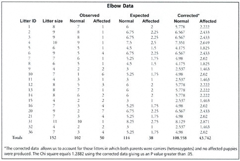
Table 1: Litters of Newfoundlands having at least 1 affected puppy in each litter and having phenotypically normal parents
With this degree of accuracy and an affected to affected mating producing at least four or five puppies, all of which should be affected if this is a simple autosomal recessive trait, will allow us to publish this data in a reviewed journal. The affected to affected mating allows us to check our family data. We expect all puppies born to be affected, and if one or more is not affected, we would call the trait polygenic or a recessive trait with incomplete penetrance.
Conclusions
At the present time, this trait does not appear to be limited to any specific line(s), but future studies may allow us to narrow down a set of dogs which need to be correctly bred to produce normal, healthy puppies and avoid this trait.
It is important to remember that the Newfoundland has 46 other genetic diseases and that this particular disorder must be put into a perspective that allows us to breed Newfoundlands which are free of any major disease, not just this one. We would like to give our profound respect to those breeders who are helping us with this project. We need to sincerely thank them, not throw a brick at them.
The Chi-Square Statistic Defined by Pat Randall, Ph.D.
This preliminary report on "elbow anomaly" shows that the data suggest an autosomal recessive mode of inheritance. The Chi-square statistic and the strategy used to test this hypothesis may be uFLA/CRHLmiliar to many readers.
Most readers will know that with an autosomal recessive disorder, breeding two heterozygotes (carriers) should, in the long run, produce about 25% affected offspring, i.e. equal numbers of AA, Aa, aA, and aa where A is the "normal" gene and a is the defective form of the gene. The aa or homozygous recessive genotype represent the affected dogs. The sires and dams of all the litters in the study are phenotypically normal (non-affected), but have produced at least 1 affected puppy in the litter. Thus, if the disorder is recessive, these would have to be carrier by carrier breedings. If the ratio of normal to affected offspring approximate our expected 3: 1 ratio, we would consider this to be evidence that it is, indeed, an autosomal recessive disorder.
The "Chi-square" statistic is a single number calculated that reflects the deviation of the observed numbers of affected and normal offspring from the numbers expected from an autosomal recessive inheritance. The greater the deviation, the larger the Chi-square value. We calculate the Chi-square and then determine the probability of getting a deviation (chi-square) this large just by chance. If that probability is small (usually less than 5%) we will, at least tentatively, reject the hypothesis that gives rise to the 3: 1 expected ratio. The Chi-square in this study is quite small and represents only a very small deviation from what we expect. Since we are only examining litters with at least one affected puppy, the expected proportion of affecteds will actually be somewhat larger than 25% since some of the normals making up the 3: 1 theoretical ratio would come from litters in which all the puppies were normal.
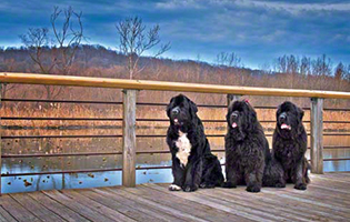
 Figure 1: Affected male puppy with bowing of the front legs and deviation of the pasterns.
 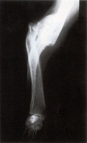 Figure 2: Anterior to Posterior x-ray view of the forelimb of an affected adult
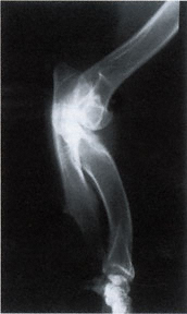 Figure 3: Lateral x-ray view of the forelimb of an affected adult
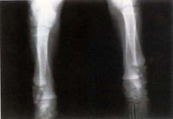 Figure 4: Anterior to posterior x-ray of the forelimbs (elbows to pasterns) of a normal puppy
 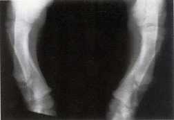 Figure 5: Anterior to posterior x-ray of the forelimbs (elbows to pasterns) of an affected puppy